Ureters, Kidney, Bladder and Urethra
Home > Clinical Concepts In Radiation Oncology > Anatomy, Physiology > Abdomen, Pelvis, GI and GU > Ureters, kidney, etc
Can we please get your advice on this one question?
The urinary system is one of the excretory systems of the body. It consists of the following structures:
• 2 kidneys, which secrete urine
• 2 ureters, which convey the urine from the kidneys to the urinary bladder
• 1 urinary bladder where urine collects and is temporarily stored
• 1 urethra through which the urine is discharged from the urinary bladder to the exterior.
Kidney:
The kidneys lie on the posterior abdominal wall, one on each side of the vertebral column, behind the peritoneum and below the diaphragm. They extend from the level of the 12th thoracic vertebra to the 3rd lumbar vertebra, receiving some protection from the lower rib cage. The right kidney is usually slightly lower than the left, probably because of the considerable space occupied by the liver. Kidneys are bean-shaped organs, about 11 cm long, 6 cm wide, 3 cm thick and weigh 150 g. They are embedded in, and held in position by, a mass of fat. A sheath of fibroelastic renal fascia encloses the kidney and the renal fat.
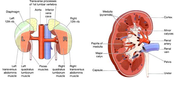
Fig: (a) Posterior view of the kidney showing the areas associated structures. Fig (b) cross section of kidney
Gross Structure of Kidney:
There are three areas of tissue which can be distinguished when a longitudinal section of the kidney is viewed with the naked eye (Fig. b)
• A fibrous capsule, surrounding the kidney
• The cortex, a reddish-brown layer of tissue immediately below the capsule and outside the pyramids
• The medulla, the innermost layer, consisting of pale conical-shaped striations, the renal pyramids.
The hilum is the concave medial border of the kidney where the renal blood and lymph vessels, the ureter and nerves enter. The renal pelvis is the funnel-shaped structure which acts as a receptacle for the urine formed by the kidney (Fig. b). It has a number of distal branches called calyces, each of which surrounds the apex of a renal pyramid. Urine formed in the kidney passes through a papilla at the apex of a pyramid into a minor calyx, then into a major calyx before passing through the pelvis into the ureter. The walls of the pelvis contain smooth muscle and are lined with transitional epithelium. Peristalsis of the smooth muscle originating in pacemaker cells in the walls of the calyces propels urine through the pelvis and ureters to the bladder. This is an intrinsic property of the smooth muscle, and is not under nerve control.
The unit of kidney is called Nephron.
Functions of the kidney:
The main functions of the kidney are,
1. formation and secretion of urine
2. production and secretion of erythropoietin, the hormone responsible for controlling the rate of formation of red blood cells
3. Production and secretion of renin, an important enzyme in the control of blood pressure.
Ureter:
The ureters are the tubes that convey urine from the kidneys to the urinary bladder (Fig. c). They are about 25 to 30 cm long with a diameter of about 3 mm.
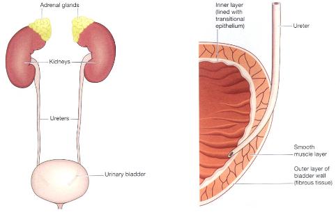
Fig.(c) shows the 2 ureters from the kidney to bladder
The ureters consist of three layers of tissue:
• An outer covering of fibrous tissue, continuous with the fibrous capsule of the kidney
• A middle muscular layer consisting of interlacing smooth muscle fibres that form a syncytium spiralling round the ureter, some in clockwise and some in anticlockwise directions and an additional outer longitudinal layer in the lower third
• An inner layer, the mucosa, lined with transitional epithelium.
Functions:
The ureters propel the urine from the kidneys into the bladder by peristaltic contraction of the smooth muscle layer. This is an intrinsic property of the smooth muscle and is not under autonomic nerve control. The waves of contraction originate in a pacemaker in the minor calyces. Peristaltic waves occur several times per minute, increasing in frequency with the volume of urine produced, and send little spurts of urine into the bladder.
Urinary Bladder:
The urinary bladder is a reservoir for urine. It lies in the pelvic cavity and its size and position vary, depending on the amount of urine it contains. When distended, the bladder rises into the abdominal cavity.
The bladder is roughly pear-shaped, but becomes more oval as it fills with urine. It has anterior, superior and posterior surfaces. The posterior surface is the base. The bladder opens into the urethra at its lowest point, the neck.
The peritoneum covers only the superior surface before it turns upwards as the parietal peritoneum, lining the anterior abdominal wall. Posteriorly it surrounds the uterus in the female and the rectum in the male.
The bladder wall is composed of three layers:
1. The outer layer of loose connective tissue, containing blood and lymphatic vessels and nerves, covered on the upper surface by the peritoneum
2. The middle layer, consisting of a mass of interlacing smooth muscle fibres and elastic tissue loosely arranged in three layers. This is called the detrusor muscle and it empties the bladder when it contracts
3. The mucosa, lined with transitional epithelium
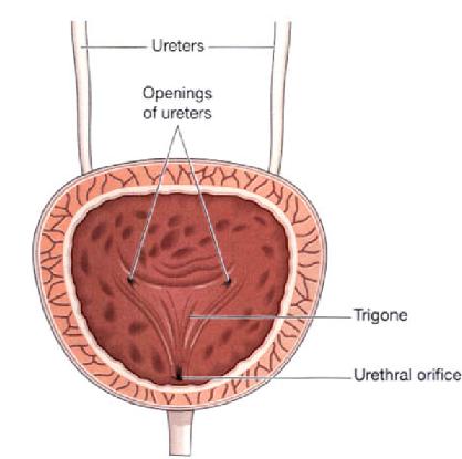
Fig: (d) shows the cross section view of urinary bladder
When the bladder is empty the inner lining is arranged in folds, or rugae, and these gradually disappear as the bladder fills. The bladder is distensible but when it contains 300 to 400 ml the awareness of the desire to urinate is initiated. The total capacity is rarely more than about 600 ml. The three orifices in the bladder wall form a triangle or trigone (Fig. d). The upper two orifices on the posterior wall are the openings of the ureters. The lower orifice is the point of origin of the urethra. Where the urethra commences is a thickening of the smooth muscle layer forming the internal urethral sphincter. This sphincter is not under voluntary control.
The female urethra is approximately 4 cm long. It runs downwards and forwards behind the symphysis pubis and opens at the external urethral orifice just in front of the vagina. The external urethral orifice is guarded by the external urethral sphincter which is under voluntary control. Except during the passage of urine, the walls of the urethra are in close apposition. The male urethra is described in detail in Chapter 19, but in both sexes the basic structure is the same. Its walls consist of three layers of tissue. the muscle layer, continuous with that of the bladder. At its origin there is the internal urethral sphincter consisting mainly of elastic tissue and smooth muscle fibres, under autonomic nerve control. Slow and continuous contraction of this sphincter keeps the urethra closed. In the middle third there is skeletal muscle surrounding the urethra, under voluntary nerve control, that forms the external urethral sphincter the submucosa, a spongy layer containing blood vessels and nerves the mucosa, which is continuous with that of the bladder in the upper part. In the lower part the lining consists of stratified squamous epithelium, continuous externally with the skin of the vulva.
Urethra:
The urethra is a canal extending from the neck of the bladder to the exterior, at the external urethral orifice. Its length differs in the male and in the female.
Questions:
1. What are the main functions of the kidney?
a) formation and secretion of urine
b) production and secretion of erythropoietin
c) Production and secretion of renin
d) All
2. What is the capacity of the urinary bladder?
a) 600ml
b) 100ml
c) 2 liters
d) None of these
Answer:
d) All
a) 600 ml
References:
1. Ross and Wilson, Anatomy and physiology in health and illness by Anne Waugh and Allison Grant.
2. Anatomy at a Glance by Omar faiz and David Moffat
3. www.wikipedia.com
Home > Clinical Concepts In Radiation Oncology > Anatomy, Physiology > Abdomen, Pelvis, GI and GU > Ureters, kidney, etc
FREE Infographic What successful people believe. What successful people do
Dictionary of Cancer Terms
Need help understanding a word? Here is an electronic resource that gives meaning to Cancer terms and their usage.

StrengthsFinder 2.0