Magnification
Factor
Home > Treatment Planning > Treatment Volume Localization > Fluoroscopic Simulation > Magnification Factors
Can we please get your advice on this one question?
Magnification:
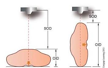
As the X-ray beam diverges the image produced on the film is always slightly larger than the original image, and this effect is called magnification.
The Magnification factor is the degree of magnification and is calculated by:
M = SID/SOD
Where: M is the magnification factor
The magnification factor permits calculation of the actual size of an object that is projected as an image by using the formula:
O = I/M
Where,
O = object size
I = image size
M = magnification factor
Then, substitute the value of M above
M = SID/SOD
O = I / (SID/SOD)
Magnification consideration while planning:
In 2D treatment planning, block apertures are defined from simulation films; therefore, accurate specification of the magnification factors is important. Three-dimensional treatment-planning systems are more complex in that apertures can be defined interactively using BEV computer displays. In this approach all volumes of interest are projected onto a plane (usually passing through the Isocenter) along ray lines that originate the source. Block apertures or MLC settings can be entered to encompass the projected target. Geometrical distortions due to programming bugs can cause serious errors in the irradiated volume due to mis-registration of the treatment fields and the target volume. The BEV accuracy as a function of gantry angle, collimator angle, field size, and isocenter distance should be confirmed prior to use and checked at a regular intervals and after any software modification.
In radiograph image depends on the distance between the patient and the film, and the distance between the patient and the focus of the x-rays. The minimum amount of magnification is produced if the film is close to the patient and the patient is at a long distance from the focus of the x-ray tube.
The magnification can be measured, which is useful in order to calculate the image size on the film relative to the size on the actual patient.
Questions:
1. If the SID is 40 inches (100 cm) and the SOD is 30inches (75 cm), what is the magnification factor?
a) 1.22
b) 1.33
c) 1.44
d) 1.55
Answer:
1. b) 1.33
References:
1. books.google.co.in/books?isbn=1439058725
2. Technical Basis of Radiation Therapy - Practical Clinical Applications__Medical Radiology___Radiation Oncology_
Home > Treatment Planning > Treatment Volume Localization > Fluoroscopic Simulation > Magnification Factors
FREE Infographic What successful people believe. What successful people do
Dictionary of Cancer Terms
Need help understanding a word? Here is an electronic resource that gives meaning to Cancer terms and their usage.

StrengthsFinder 2.0