Imaging Studies

Home > Clinical Concepts In Radiation Oncology > Patient Evaluation > Imaging Studies
Can we please get your advice on this one question?
X-ray Imaging:
X-ray imaging is perhaps the most familiar type of imaging. Images produced by X-rays are due to the different absorption rates of different tissues. Calcium in bones absorbs X-rays the most, so bones look white on a film recording of the X-ray image, called a radiograph. Fat and other soft tissues absorb less, and look gray. Air absorbs least, so lungs look black on a radiograph. The most familiar use of X-rays is checking for broken bones, but X-rays are also used in cancer diagnosis. For example, chest radiographs (fig:a)and mammograms are often used for early cancer detection or to see if cancer has spread to the lungs or other areas in the chest. Mammograms use X-rays to look for tumors or suspicious areas in the breasts.
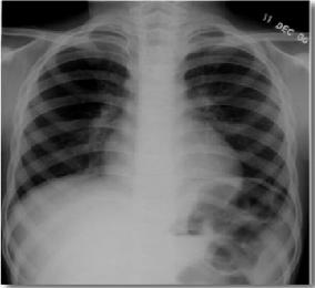
Fig: a shows the typical x-ray radiograph
Mammography:
Conventional mammography uses X-rays to look for tumors or suspicious areas in the breasts. Digital mammography also uses X-rays, but the data is collected on computer instead of on a piece of film. This means that the image can be computer-enhanced, or areas can be magnified. Eventually, a computer could in certain appropriate situations, screen digital mammograms, theoretically detecting suspicious areas that human error might miss.
Ct scan:
A computed tomography scan (CT scan, also called a CAT scan) uses computer-controlled X-rays to create images of the body. However a radiograph and a CT scan show different types of information. Although an experienced radiologist can get a sense for the approximate three-dimensional location of a tumor from a radiograph, in general, a plain radiograph is two-dimensional.
An arm or chest radiograph looks all the way through a body without being able to tell how deep anything is. A CT scan is three-dimensional. By imaging and looking at several three-dimensional slices of a body (like slices of bread) a doctor could not only tell if a tumor is present, but roughly how deep it is in the body. A CT scan can be three dimensional because the information about how much of the X-rays are passing through a body is collected not just on a flat piece of film, but on a computer.
The data from a CT scan can be enhanced to be more vivid than a plain radiograph. For both plain radiographs and CT scans, the patient may be given a contrast agent to drink and/or by injection to more clearly show the boundaries between organs or between organs and tumors.
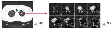
Fig:b (Left) A 1 mm spiral CT "slice" through the mid-chest region, showing both lungs. The white spot in the right lung is a suspicious nodule that could be biopsied to see if it is cancerous. (Right) A close-up view of 8 "slices" focused on a lung nodule. Compared with a traditional X-ray, a series of CT images gives the radiologist a much better sense of nodule size and its potential threat. Images courtesy of A. P. Reeves, Cornell University.
Magnetic Resonance Imaging (MRI):
Magnetic Resonance Imaging (MRI) uses radio waves in the presence of a strong magnetic field that surrounds the opening of the MRI machine where the patient lies to get tissues to emit radio waves of their own.
Different tissues (including tumors) emit a more or less intense signal based on their chemical makeup, so a picture of the body organs can be displayed on a computer screen. Much like CT scans, MRI can produce three-dimensional images of sections of the body, but MRI is sometimes more sensitive than CT scans for distinguishing soft tissues.

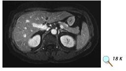
Fig:c (Top) MRI scan without contrast showing possible tumor in the liver. (Bottom) MRI scan of the same patient using contrast. Images courtesy of Dr. Peter Choyke, Clinical Center, NIH.
Questions:
1.Mammography is used for imaging the,
a) Brain
b) Chest
c) Breast
d) Liver
2. What is the principle behind in the x-ray image formation?
a) Reflection
b) Differential absorption
c) Scattering
d) All
Answers:
c) Breast
b) Differential absorption
References:
1. http://imaging.cancer.gov
3. http://www.elsevierscitech.com
4. http://8cs25.edu.glogster.com
Home > Clinical Concepts In Radiation Oncology > Patient Evaluation > Imaging Studies
FREE Infographic What successful people believe. What successful people do
Dictionary of Cancer Terms
Need help understanding a word? Here is an electronic resource that gives meaning to Cancer terms and their usage.

StrengthsFinder 2.0